Visual Impairments
By: Hannah Hunt OTR/L
Clinical Condition Overview
Low Vision
The National Eye Institute defines low vision as a condition that cannot be corrected with “eyeglasses, contacts, or other standard treatments like medicine or surgery” and is severe enough to interfere with daily activities (National Eye Institute, 2020). Low vision is categorized according to the World Health Organization as the best corrected visual acuity of 20/70 in the better seeing eye (Low Vision and Vision Rehabilitation, n.d.). The most common diseases causing vision loss in the aging population are age-related macular degeneration, glaucoma, cataract, and diabetic retinopathy (Quillen, 1999).
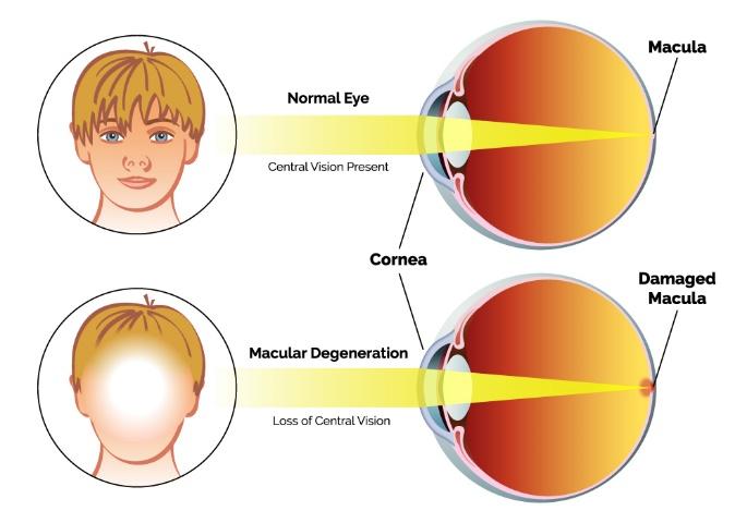
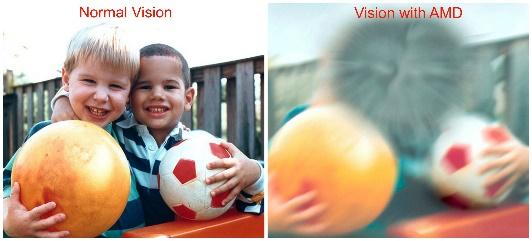
- Age-Related Macular degeneration (AMD) is a disease that causes progressive deterioration of the cells in the light sensitive tissue in the back of the eye known as the retina. AMD affects only the photoreceptors that make up the center of the retina, known as the macula, and is characterized by a loss of sharp, detailed vision in the central visual field (National Eye Institute, 2019). AMD is classified into two types: dry and wet.
Reference: https://naplesoptical.com/what-is-macular-degeneration/
Dry vs Wet AMD: In the most common form, known as non-exudative or “dry” AMD, vision loss is typically slow progressing. Dry macular degeneration occurs when parts of the macula become deteriorate and a yellowish protein called drusen grow and deposit beneath the retina. As this develops, central vision gradually worsens.
As this disease progresses, abnormal blood vessels develop under the macula in reaction to the damaged cells, known as “wet” macular degeneration. If these blood vessels break and bleed onto the macula, sudden vision loss can occur (Low Vision and Vision Rehabilitation, n.d.).
Characteristics: AMD results in the loss in the ability to see fine detail where central vision may become wavy or blurred. AMD may result in blind spots known as scotomas, and in advanced cases, central vision may be completely lost. While peripheral vision is preserved, individuals may also experience visual distortion, poor visibility in low light conditions, reduced contrast sensitivity function, reduced color vision, reduced dark/light adaptation, and photosensitivity (Warren & Barstow, 2011, p.28). Reduced contrast sensitivity often causes may cause difficulty distinguishing items of similar color, difficulty recognizing faces, and seeing curbs or steps. Individuals with AMD often benefit from magnification for reading due to reduced central acuity.
Reference: https://www.scottsdaleeye.com/macular-degeneration-need-know/
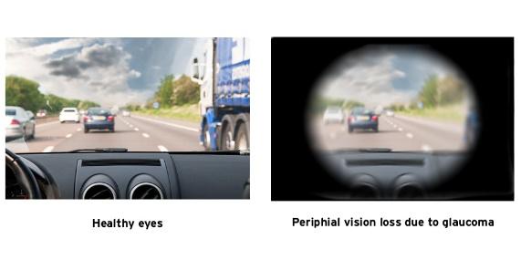
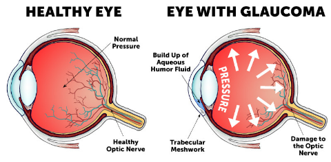
- Glaucoma is a group of eye conditions in which the optic nerve is damaged from increased pressure inside the eye. Glaucoma is characterized by peripheral vision loss, or tunnel vision, and can lead to total blindness if left untreated (Warren & Barstow, 2011, p. 41).
Reference: https://www.aoa.org/healthy-eyes/eye-and-vision-conditions/glaucoma?sso=y
- Etiology: When fluid in the anterior chamber of the eye becomes blocked, intraocular pressure can build causing damage to the optic nerve. The most prevalent form of glaucoma, called primary open-angle glaucoma, is chronic and progresses slowly over time. However, glaucoma can be congenital, age-related, or traumatic (Glaucoma, n.d).
Reference: https://www.wolfeeyeclinic.com/medical-services/glaucoma
- Diabetic retinopathy is an eye disease that can cause varying patterns of vision loss in individuals with diabetes. DR is a continuum pathology that begins with a form known as non-proliferative or background diabetic retinopathy.
- Non-proliferative and proliferative DR: High levels of glucose in the blood can destroy the integrity of the blood vessels of the retina, reducing the amount of oxygen delivered to the retinal cells. These damaged vessels can begin to leak fluid onto the surface of the retina resulting in visual field loss. As the disease progresses, the retina responds to the lack of blood supply by developing new blood vessels along its surface called neovascularization. This is known as the proliferative form of diabetic retinopathy. If these abnormal blood vessels rupture, fluid can leak into the vitreous of the eye and may result in the individual seeing dark, floating spots, and in some cases, experience severe and sudden vision loss.
- Characteristics: Persons with DR may have scattered spotty areas of vision loss called scotomas. If a complication known as macular edema occurs where damaged blood vessel leak fluid in the macula, central vision can be severely impaired. Other common characteristics include decreased contrast sensitivity function, reduced color discrimination, poor night vision, and fluctuations in vision (Warren & Barstow, 2011, p.34-35).
- Cataract is the leading cause of blindness worldwide (Quillen, 1999). Cataract is characterized by opacification or cloudiness of the lens of the eye that interfere with vision function (Warren & Barstow, 2011, p.36). Patients with cataracts may experience blurred vision, glare, reduced contrast sensitivity function, and reduced color perception. Cataract progression is typically slow, with gradual loss of vision over months to years. However, some types of cataract progress more rapidly. Cataract surgery is a procedure in which the lens of the eye is removed and, in most cases, replaced it with an artificial lens. Cataract surgery is an outpatient procedure performed under local or topical anesthesia with rare instance of complications. This surgery should be considered when the cataract reduces vision function to a level that interferes with everyday activities (Warren & Barstow, 2011, p. 36).
Vision Loss related to neurological conditions
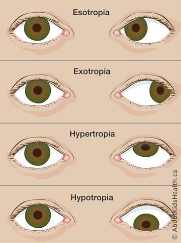
Brain injury, stroke, and tumors can result in a variety of conditions impacting visual perception. Some of these disturbances are caused by “direct trauma to the orbital content, cranial nerves and other brain areas” (Cohen, 1989). Neurological conditions and trauma may impact the six extraocular muscles that control the eyes resulting in a condition known as strabismus where the eyes do not align while looking at an object. Strabismus may cause conditions known as exotropia, esotropia, hypertropia, and hypotropia.
6 Extraocular muscles: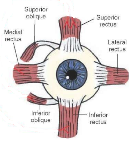
Strabismus:
Reference: https://www.aboutkidshealth.ca/Article?contentid=836&language=English
Cranial Nerve Palsies:
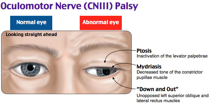
- Oculomotor nerve palsy (3rd cranial nerve palsy) is a condition resulting from damage to the oculomotor nerve. Oculomotor nerve palsy typically results in a characteristic displacement outward (exotropia) and downward (hypotropia) due to paralysis of the medial rectus, superior rectus, inferior recuts, and inferior oblique muscles. The affected individual will also have a ptosis, or drooping of the eyelid and mydriasis (pupil dilation). Common symptoms may include closing of one eye, abnormal head posture such as tilting or head turning, reports of blurry or double vision, over or under-shooting when reaching for objects, and poor depth perception.
Reference: https://mobilephysiotherapyclinic.in/oculomotor-nerve/
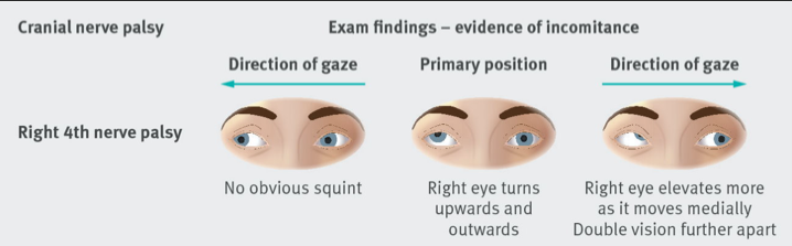
- Trochlear nerve palsy (4th cranial nerve palsy) results in a weakening or paralysis of the superior oblique muscle where the eye tends to drift upwards. The resultant vertical strabismus is termed a “hypertropia”. Adults who have a fourth nerve palsy will frequently observe double vision and head tilt to the side opposite the palsied eye, a natural compensation for the vertical strabismus.
Reference: https://eyesoneyecare.com/resources/ods-guide-diagnosis-and-management-cranial-nerve-iv-palsy/
- Abducens Nerve Palsy (6th cranial nerve palsy): is a disorder affecting the abducens cranial nerve controlling the lateral rectus muscle. This results in an esotropia, with the affected eye turning in medially towards the nose. Common symptoms include double vision when looking side to side and increased head movement in order to reduce double vision.
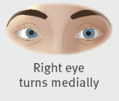
Reference: https://eyesoneyecare.com/resources/ods-guide-diagnosis-and-management-cranial-nerve-iv-palsy/
- Treatment of cranial nerve palsies may include: eye patching, prism therapy, vision rehabilitation therapy, and/or surgical intervention.
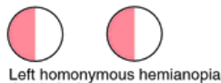
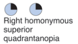
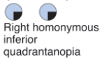
- Hemianopia and quadranopia are types of visual field loss caused by stroke, traumatic brain injury, tumor, and other neurological conditions. Symptoms of these types of field loss include a narrowed search pattern confined to the sound side, slowed visuals scanning to the affected visual field, trouble locating items on the blind side, and missing and/or mis-identifies visual detail on the blind side. Individuals with visual field loss may demonstrate difficulty with navigation, self-care, and reading. Intervention may include visual scanning training focused on complete and organized visual search from left to right, increasing visual search speeds to quickly obtain information needed from the environment. This may include use of technology such as the Dynavision Light Board, narrated walks (avoiding objects/hazards), treasure hunts (finding targets/things), and large table top activities focused on encouraging visual scanning.

- Hemi-inattention/Neglect– visual inattention or neglect is a deficit of visual perception. Common symptoms of neglect may include: missing or bumping into objects on one side during ambulation, recognizing only one half of the environment. Testing for neglect may include Star Cancellation, Line Bisection, Figure Copying Test, Clock Drawing Test, and the Catherine Bergego Scale.
- Treatment of neglect may include:
- increasing awareness of the neglected visual field
- visual scanning training or increasing ability to scan/head turn towards the affected side
- adding brightly colored tape on the neglected side of an object, such as a clock, door threshold, or page of a book
- the use of yoked prisms
- increasing proprioception on the neglected side of the body.
- Treatment of neglect may include:
- Photosensitivity following TBI can also impact ADL performance and require filtered lens and/or managing of light sources both indoors and outdoors.
References:
Cohen, M., Groswasser, Z., Barchadski, R., & Appel, A. (1989). Convergence insufficiency in brain-
injured patients. Brain injury, 3(2), 187–191. https://doi.org/10.3109/02699058909004551
Glaucoma. (n.d.). American Optometric Association. Retrieved January 23, 2022, from https://www.aoa.org/healthy-eyes/eye-and-vision-conditions/glaucoma?sso=y
Quillen D. A. (1999). Common causes of vision loss in elderly patients. American family
physician, 60(1), 99–108. https://www.aafp.org/afp/1999/0701/p99.html
Low Vision and Vision Rehabilitation. (n.d.). American Optometric Association. Retrieved January 23, 2022, from https://www.aoa.org/healthy-eyes/caring-for-your-eyes/low-vision-and-vision-rehab?sso=y
National Eye Institute. (2019). Age-Related Macular Degeneration | National Eye Institute. Nih.gov. https://www.nei.nih.gov/learn-about-eye-health/eye-conditions-and-diseases/age-related-macular-degeneration
National Eye Institute. (2020). Low Vision. https://www.nei.nih.gov/learn-about-eye-health/eye-conditions-and-diseases/low-vision
Warren, M., Barstow, E. A., & American Occupational Therapy Association. (2011). Occupational Therapy Interventions for Adults with Low Vision. Amsterdam University Press.